Elektrine
Log in Register
Paige Chat Timeline Gallery Friends Email Drive DNS Private DNS Domains VPN Kairo Nerve
Remote

StructBiolCommunications

@ActaCrystF@mstdn.science
mastodon 4.6.8
  • Open on mstdn.science

Acta Crystallographica Section F: Structural Biology Communications is a structural biology communications journal published by the IUCr

0 Followers
0 Following
20 Posts
Joined December 22, 2022
PGP:
https://keyoxide.org/hkp/0ADA8BF24CC573A3FF2CD1F0B96A542ABBA76EBF
Web:
http://journals.iucr.org/f
Open post
StructBiolCommunications @ActaCrystF@mstdn.science
· 7mo ago

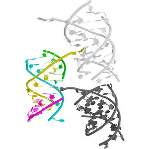
This structure of a tRNA acceptor-stem mimic demonstrates the use of bromine and phosphorus anomalous scattering for RNA structure determination #Crystallography #AnomalousScattering #NucleicAcids https://doi.org/10.1107/S2053230X26000658

Crystal structure of a tRNA acceptor-stem mimic at 1.94 Å resolution
Acta Crystallographica Section F

Crystal structure of a tRNA acceptor-stem mimic at 1.94 Å resolution

This paper reports the structure solution of an RNA complex using anomalous scattering.

1
0
1
0
Open post
StructBiolCommunications @ActaCrystF@mstdn.science
· 5mo ago

New issue is out! https://journals.iucr.org/f/issues/2026/05/00/

On the cover: the atomic structure of human dystrophin spectrin-like repeat 24 determined at 2.14 Å resolution https://journals.iucr.org/f/issues/2026/05/00/rl5207/index.html

journals.iucr.org

(IUCr) Acta Crystallographica Section F Volume 82, Part 5, May 2026

Acta Crystallographica Section F Structural Biology Communications

0
0
1
0
Open post
StructBiolCommunications @ActaCrystF@mstdn.science
· 5mo ago

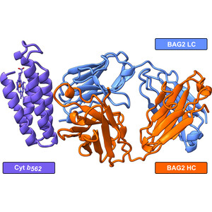
Modifying the bacterial strain led to cleaner bacterial expression of a synthetic BRIL antibody that facilitates protein structure determination via both crystallography and cryo-EM #CryoEM #XRayCrystallography #BRIL https://doi.org/10.1107/S2053230X26001548
Optimized bacterial expression of a synthetic BRIL antibody
Acta Crystallographica Section F

Optimized bacterial expression of a synthetic BRIL antibody

Here, we document the generation of a T7 Express ΔcybC strain allowing contaminant-free expression of the anti-BRIL Fab BAG2. We also report the crystal structure of BAG2 in complex with native cytochrome b562, a complex arising from expression in canonical Escherichia coli strains.

0
0
0
0
Open post
StructBiolCommunications @ActaCrystF@mstdn.science
· 5mo ago

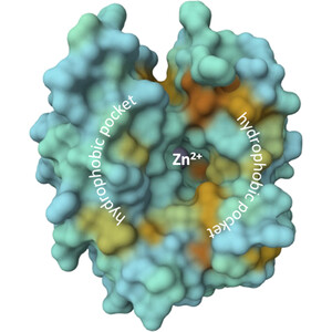
High-resolution structures of α-bCA2 with sulfonamide derivatives provides experimentally supported information defining their binding mode and supporting inhibitor optimization #CarbonicAnhydraseII #SulfonamideInhibitors #XRayCrystallography https://doi.org/10.1107/S2053230X26002128
X-ray analysis of complexes of carbonic anhydrase II with 1,3-oxazole-containing sulfonamide derivatives elucidates the structural basis for their exceptionally high inhibitory potency
Acta Crystallographica Section F

X-ray analysis of complexes of carbonic anhydrase II with 1,3-oxazole-containing sulfonamide derivatives elucidates the structural basis for their exceptionally high inhibitory potency

High-resolution X-ray structures of carbonic anhydrase II in complex with two 1,3-oxazole-containing sulfonamide inhibitors define the interactions responsible for their exceptional inhibitory potency and reveal how subtle ligand instability can influence electron-density interpretation and model refinement.

0
0
0
0
Open post
StructBiolCommunications @ActaCrystF@mstdn.science
· 5mo ago

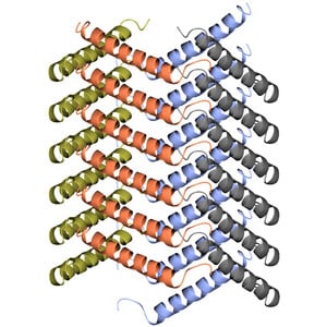
An acylated GLP-1/GIP analogue peptide forms a uniquely porous spiral crystal structure composed of clockwise-ascending monomers in a square pattern #GLP1 #Peptides #Crystallization https://doi.org/10.1107/S2053230X26001937
Crystallization and 1.6 Å resolution crystal structure of an acylated GLP-1/GIP analogue peptide
Acta Crystallographica Section F

Crystallization and 1.6 Å resolution crystal structure of an acylated GLP-1/GIP analogue peptide

The 1.6 Å resolution diffraction and subsequent structure solution of an acylated GLP-1/GIP analogue lipopeptide is reported. This represents the first published data on the crystal structure of an unbound GLP-1 and/or GIP analogue lipopeptide.

0
0
0
0
Open post
StructBiolCommunications @ActaCrystF@mstdn.science
· 5mo ago

A comprehensive survey catalogues all pentatricopeptide repeat protein crystal structures, analysing their crystallographic properties, structural features and contributions to understanding function #PentatricopeptideRepeatProteins #RNARecognition https://doi.org/10.1107/S2053230X26002311
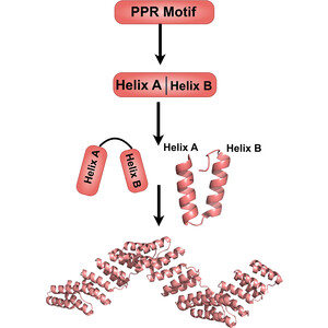
Structures of pentatricopeptide repeat proteins
Acta Crystallographica Section F

Structures of pentatricopeptide repeat proteins

A number of crystal, cryo-EM and solution structures of pentatricopeptide repeat proteins have been solved, revealing details of how they interact with single-stranded RNA and other proteins, as well as their conformational repertoire.

0
0
0
0
Open post
StructBiolCommunications @ActaCrystF@mstdn.science
· 6mo ago

Our April issue is out now! https://journals.iucr.org/f/issues/2026/04/00/

Crystallization was investigated as a preparative strategy for a GLP-1/GIP lipopeptide and led to what is believed to be the first reported crystal structure of an unbound GLP-1/GIP analogue https://doi.org/10.1107/S2053230X26001937

journals.iucr.org

(IUCr) Acta Crystallographica Section F Volume 82, Part 4, April 2026

Acta Crystallographica Section F Structural Biology Communications

0
0
0
0
Open post
StructBiolCommunications @ActaCrystF@mstdn.science
· 6mo ago

The structure of endo-β-N-acetylglucosaminidase was determined at 1.8 Å resolution, revealing a unique spatial arrangement of domain IV, which is suggested to contribute to substrate recognition #EndoNAcetylglucosaminidases #GH85 #ComplexNGlycans https://doi.org/10.1107/S2053230X26001214
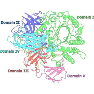
Crystal structure of endo-β-N-acetylglucosaminidase HSα
Acta Crystallographica Section F

Crystal structure of endo-β-N-acetylglucosaminidase HSα

Structural analysis of the GH85 enzyme endo-β-N-acetylglucosaminidase HSα reveals a novel domain that is potentially involved in complex N-glycan recognition.

0
0
0
0
Open post
StructBiolCommunications @ActaCrystF@mstdn.science
· 6mo ago

The structure of a chimeric efflux pump was determined by cryoEM under apo and kanamycin-supplemented conditions and adopted symmetric-like and asymmetric conformations #PseudomonasAeruginosa #DrugEffluxTransporters #MembraneProteins https://doi.org/10.1107/S2053230X26001202
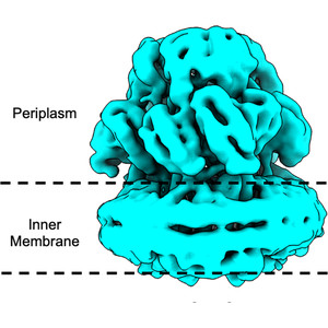
Cryo-EM structures of a MexB–MexY chimeric efflux pump reveal that large open clefts are intrinsic to the MexY porter domain
Acta Crystallographica Section F

Cryo-EM structures of a MexB–MexY chimeric efflux pump reveal that large open clefts are intrinsic to the MexY porter domain

In this study, cryo-EM structures of the chimeric RND transporter MexBYB, composed of MexB and MexY, are presented, revealing distinct symmetric-like and asymmetric conformations under apo and drug-supplemented conditions. These structures show that the unusually large open clefts observed in MexBYB, similar to those in MexY, represent a unique conformational feature of the MexY porter domain.

0
0
0
0
Open post
StructBiolCommunications @ActaCrystF@mstdn.science
· 6mo ago

MBP fusion is an effective and generalizable approach to enable crystallographic studies of engineered binding proteins that are otherwise difficult to crystallize #Monobody #Aggregation #MaltoseBindingProtein https://doi.org/10.1107/S2053230X26000798
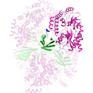
Crystallization and X-ray structure of a highly aggregation-prone monobody engineered for high-affinity small-molecule recognition
Acta Crystallographica Section F

Crystallization and X-ray structure of a highly aggregation-prone monobody engineered for high-affinity small-molecule recognition

This report describes the crystallization and X-ray structure of a highly aggregation-prone monobody engineered for small-molecule recognition. By employing a maltose-binding protein fusion strategy with optimized inter-domain linkers, we successfully obtained diffraction-quality crystals of the protein–ligand complex and determined its structure.

0
0
0
0
Open post
StructBiolCommunications @ActaCrystF@mstdn.science
· 7mo ago

This report of the structure of a carbon-nitrogen hydrolase https://doi.org/10.1107/S2053230X26001330 is the first contribution to the Early Career Scientists in Structural Science special issue! Find out more, join in and submit at https://journals.iucr.org/early-career/special-issue-2026.html
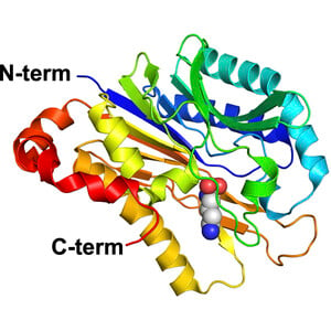
Crystal structure of carbon–nitrogen hydrolase from Helicobacter pylori G27
Acta Crystallographica Section F

Crystal structure of carbon–nitrogen hydrolase from Helicobacter pylori G27

H. pylori carbon–nitrogen hydrolase was produced and crystallized, and its 2.1 Å resolution structure is reported.

0
0
0
0
Open post
StructBiolCommunications @ActaCrystF@mstdn.science
· 7mo ago

Here's our March issue: https://journals.iucr.org/f/issues/2026/03/00/

On the cover: the structure of the chimeric RND-type multidrug-efflux pump protein MexBYB was determined by cryoEM under apo and kanamycin-supplemented conditions https://journals.iucr.org/f/issues/2026/03/00/ek5041/index.html

journals.iucr.org

(IUCr) Acta Crystallographica Section F Volume 82, Part 3, March 2026

Acta Crystallographica Section F Structural Biology Communications

0
0
0
0
Open post
StructBiolCommunications @ActaCrystF@mstdn.science
· 7mo ago

Sample preparation from α- and β-crustacyanin pigments purified from ex vivo material from the American lobster Homarus americanus for X-ray crystallography and cryo-electron microscopy studies #Astaxanthin #Crustacyanin #Absorption https://doi.org/10.1107/S2053230X2600018X
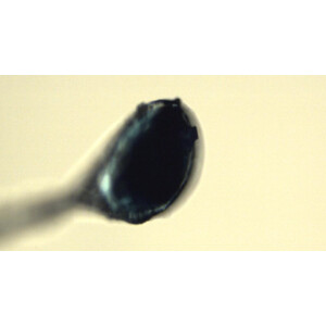
Sample purification and characterization of the α- and β-crustacyanin pigments from the American lobster for crystallographic and cryo-EM studies
Acta Crystallographica Section F

Sample purification and characterization of the α- and β-crustacyanin pigments from the American lobster for crystallographic and cryo-EM studies

The striking blue colour of the American lobster is caused by proteins that interact with the red pigment astaxanthin, causing its colour change. We have purified and characterized the pigments from the American lobster for crystallographic and cryo-EM studies.

0
0
0
0
Open post
StructBiolCommunications @ActaCrystF@mstdn.science
· 7mo ago

The highest resolution DNA-free TaXPD structure to date uniquely resolves loop 510–514 in the long-form, DNA-free TaXPD model #Helicases #Archaea https://doi.org/10.1107/S2053230X26000105
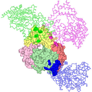
A new crystal form of the DNA-free full-length XPD helicase from Thermoplasma acidophilum
Acta Crystallographica Section F

A new crystal form of the DNA-free full-length XPD helicase from Thermoplasma acidophilum

We present a 2.13 Å resolution structure of DNA-free XPD from T. acidophilum in a novel orthorhombic lattice obtained under high ionic strength conditions. Compared with prior hexagonal forms, this structure features fewer crystal contacts and uniquely resolves loop 510–514. Interestingly, Tyr425 adopts an inward-facing conformation that would clash with DNA, suggesting a possible autoinhibitory role. These findings highlight how crystallization conditions influence lattice stability and reveal

0
0
0
0
Open post
StructBiolCommunications @ActaCrystF@mstdn.science
· 7mo ago

Potential 14-3-3 binding sites in sirtuins were identfied and the interactions of peptides representing phosphorylation sites in sirtuin 3 and sirtuin 1 with 14-3-3 proteins were characterized #Sirtuin #Phosphopeptides #Regulation https://doi.org/10.1107/S2053230X25010908
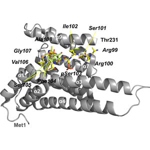
Potential 14-3-3 binding sites in sirtuins reveal extended phosphosite-recognition modes
Acta Crystallographica Section F

Potential 14-3-3 binding sites in sirtuins reveal extended phosphosite-recognition modes

Identification, binding measurements and structure determination of human sirtuin phosphopeptides in complex with human adapter protein 14-3-3σ reveal variations among tight binders and suggest extended binding modes.

0
0
1
0
Open post
StructBiolCommunications @ActaCrystF@mstdn.science
· 8mo ago

The flexible loop in Methanococcus jannaschii dihydroorotase with substrate bound adopts both the loop-out and loop-in conformations #Dihydroorotase #MethanococcusJannaschii #PyrimidineBiosynthesis https://doi.org/10.1107/S2053230X25010556
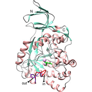
Crystal structure of Methanococcus jannaschii dihydroorotase with substrate bound
Acta Crystallographica Section F

Crystal structure of Methanococcus jannaschii dihydroorotase with substrate bound

We present the 1.87 Å resolution structure of M. jannaschii dihydroorotase complexed with the substrate carbamoyl aspartate at room temperature. Contrary to expectations, a flexible loop which is important in catalysis and closes upon substrate binding in order to stabilize the substrate and transition state is observed in both the loop-out and loop-in conformations, with the loop-out conformation having higher occupancy.

0
0
0
0
Open post
StructBiolCommunications @ActaCrystF@mstdn.science
· 8mo ago

The crystal structure of the class IIIb chitinase from Crocus vernus is reported, together with in silico analyses of inhibitor interactions and antifungal and chitinase assays #CrocusVernus #Chitinase #TIM https://doi.org/10.1107/S2053230X25009720
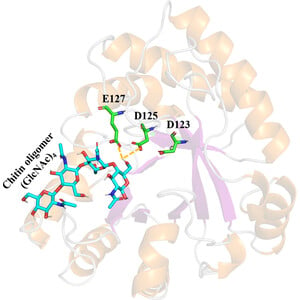
Crystal structure of Crocus vernus class IIIb chitinase: in silico ligand-binding studies and in vitro antifungal assay
Acta Crystallographica Section F

Crystal structure of Crocus vernus class IIIb chitinase: in silico ligand-binding studies and in vitro antifungal assay

This article describes the structural characterization of the class IIIb chitinase from Crocus vernus bulbs. Antifungal and chitinase assays have also been performed to evaluate the potential of the enzyme.

0
0
0
0
Open post
StructBiolCommunications @ActaCrystF@mstdn.science
· 8mo ago

Single surface substitutions can mprove the crystallizability and diffraction properties of a flexible two-domain protein even if they increase the entropy of the residue #MultidomainProtein #PointMutation #ProteinCrystallization https://doi.org/10.1107/S2053230X25010416
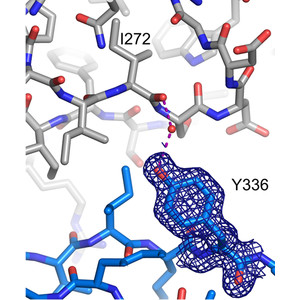
Single mutations to tyrosine or glutamate improve the crystallizability and crystal diffraction properties of a flexible two-domain protein
Acta Crystallographica Section F

Single mutations to tyrosine or glutamate improve the crystallizability and crystal diffraction properties of a flexible two-domain protein

In its wild-type form, the flexible two-domain protein InlB392 yielded poorly reproducible crystals that diffracted to low resolution. Single surface substitutions to tyrosine (T336Y) or glutamate (V333E) yielded well diffracting crystals, suggesting that surface properties, rather than interdomain flexibility, might be the primary impediment to crystallization of the wild type.

0
0
0
0
Open post
StructBiolCommunications @ActaCrystF@mstdn.science
· 8mo ago

What are the effects of the presence of polyethylene glycol in macromolecular crystals? #PolyethyleneGlycol #Mechanism #Crystallization https://doi.org/10.1107/S2053230X2501043X
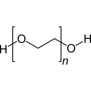
A note on the appearance of PEG in macromolecular crystals
Acta Crystallographica Section F

A note on the appearance of PEG in macromolecular crystals

The use of polyethylene glycol in the crystallization of biological macromolecules and its appearance in the resulting crystals is discussed.

0
0
0
0
Open post
StructBiolCommunications @ActaCrystF@mstdn.science
· 8mo ago

February issue now available: https://journals.iucr.org/f/issues/2026/02/00/

The cover shows the crystal structure of an RNA complex composed of four strands determined by the complementary use of bromine and phosphorus anomalous signals. Find out more at https://doi.org/10.1107/S2053230X26000658

journals.iucr.org

(IUCr) Acta Crystallographica Section F Volume 82, Part 2, February 2026

Acta Crystallographica Section F Structural Biology Communications

0
0
1
0
Back
313k7r1n3
Elektrine

Tor hidden service

elekhj7afj4qnrr4yd3bkzslsyo5jgfxw3orgjkhlcxifueodybyiiad.onion

I2P eepsite

j6b6cyk6gjmepjih7jjadxgxvvf3lzzujljuu2v4biemzpg3naya.b32.i2p

Platform

  • Email
  • Chat
  • Timeline
  • VPN
  • DNS

Company

  • About
  • Pricing
  • Contact
  • FAQ
  • Lite (no JS)

Legal

  • Terms of Service
  • Privacy Policy
  • Transparency Report
  • Report Abuse
  • Warrant Canary
  • VPN Policy

Support

  • support@elektrine.com
  • Report Security Issue
Mail client setup IMAP mail.elektrine.com:993 POP3 mail.elektrine.com:995 SMTP mail.elektrine.com:465
© 2026 Elektrine. All rights reserved. Server: 09:38:05 UTC